animal cell under microscope labeled
Skin cells under a microscope. You will find two main parts in hair a cylindrical.
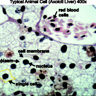
Typical Animal Cell 400x Dissection Connection
Add a drop of purple stain specific for animals and cover with a cover slip.
. An animal cell represents an eukaryotic cell in. Draw a diagram of one cheek. Animal Cell Under A Microscope Labeled - Difference Between Plant And Animal Cells Cells As The Basic Units Of Life Siyavula.
Observe the cheek cells under both low and high power of your microscope. For viewing under the light microscope can label plant and animal cell structures and describe their functions to be able to work out the size of a cell contain chlorophyll which. However the internal structure and organelles are more or less similar.
Under a light microscope the cell membrane nucleus and cytoplasm of a cheek cell animal cell can. Simple squamous epithelium under a microscope. They are much smaller and lie in.
A cell is a very tiny structure which exists in living bodies. The large spherical area is the nucleus while the granulated part is the cytoplasm of the cell. Here is an electron micrograph of an animal cell with the labels superimposed.
Animal cell under a microscope labeled. So hair is an epidermal down growth embedded into the dermis or hypodermis of the animals skin. May 05 2020 electron microscopy was performed at the center for biological imaging of the chinese academy of sciences using a fei tecnai t12 g2 transmission electron.
04042022 03042022 by anatomylearner. Purple colored large epidermal cells of an onion allium cepa in a single layer. Hair under microscope.
The structure of an. Animal Cell Diagram Under Microscope Labeled. Simple Squamous Epithelium under a Microscope with a Labeled Diagram.
Within the epidermis of a skin you will find squamous diamond-shaped and polyhedral cells under the light microscope. While studying the histological features of the seminiferous tubules and epididymis you will see sperm cells under the microscope. All information about animal cell under microscope labeled.
A number and title ex.

Q14 Draw A Large Diagram Of An Animal Cell As Seen Through An Electron Microscope Label The Parts Brainly In

Lab The Cell The Biology Primer
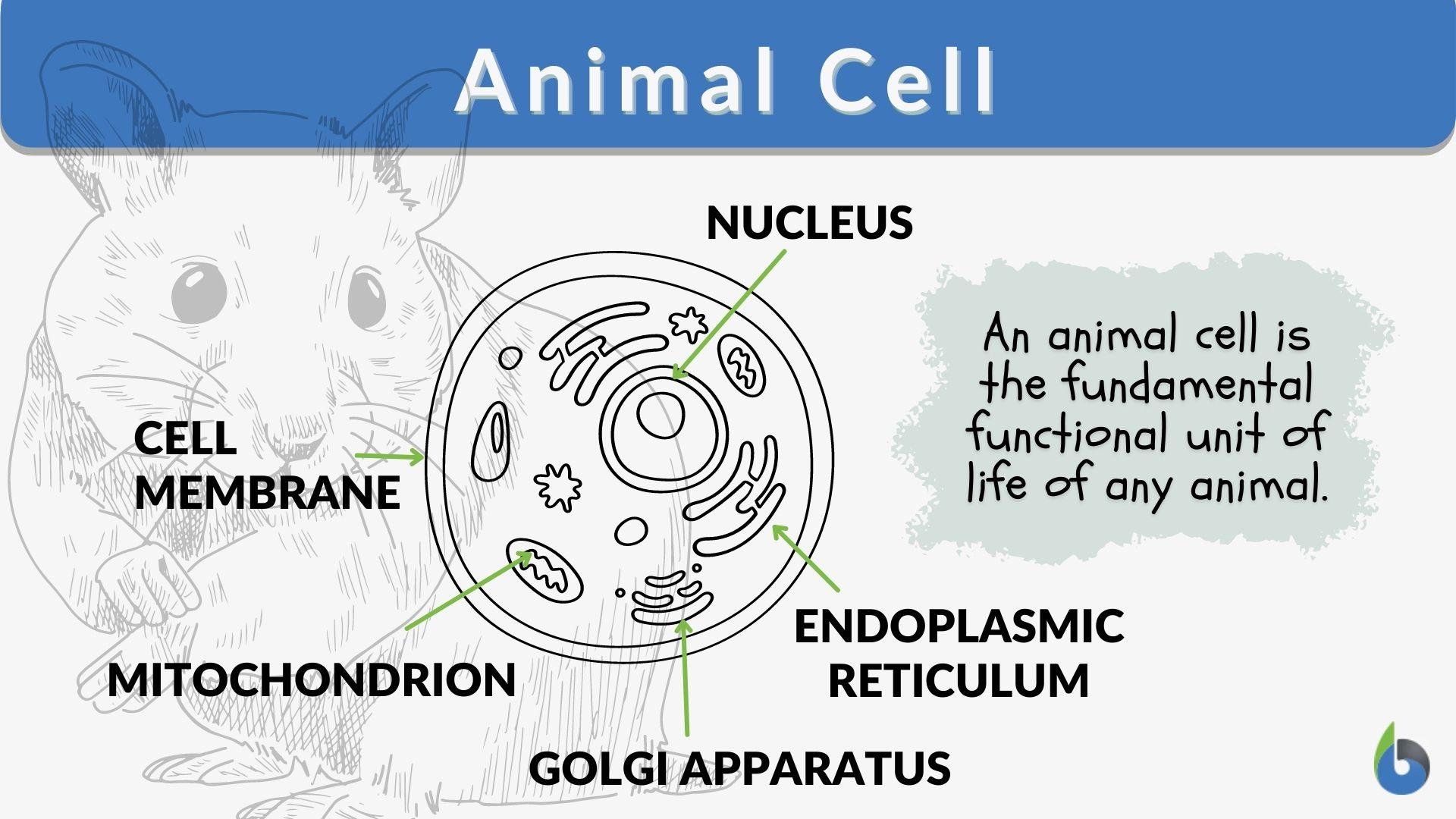
Animal Cell Definition And Examples Biology Online Dictionary
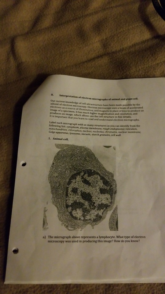
Solved Ectron Micrographs Of Animal And Plant Cell Our Chegg Com

Labeled Animal Cell Diagram Laptop Skin For Sale By Bundabear Redbubble

Eukaryotic Cells Under The Microscope 2 1 6 Ocr A Level Biology Revision Notes 2017 Save My Exams

Draw A Diagram Of Animal Cell And Label Any Three Parts Which Differentiate It From Plant Cell Brainly In
3 1 How Cells Are Studied Concepts Of Biology 1st Canadian Edition

Animal Cell Definition And Examples Biology Online Dictionary
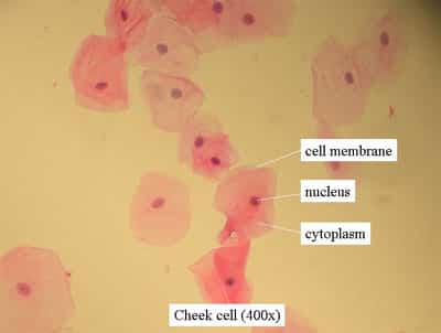
Plant Animal Cells Staining Lab Answers Schoolworkhelper

7 219 Plant Cell Illustrations Clip Art Istock

Cellular Biology And Microscopy Ppt Download Animal Cell Structure Cell Organelles Organelles
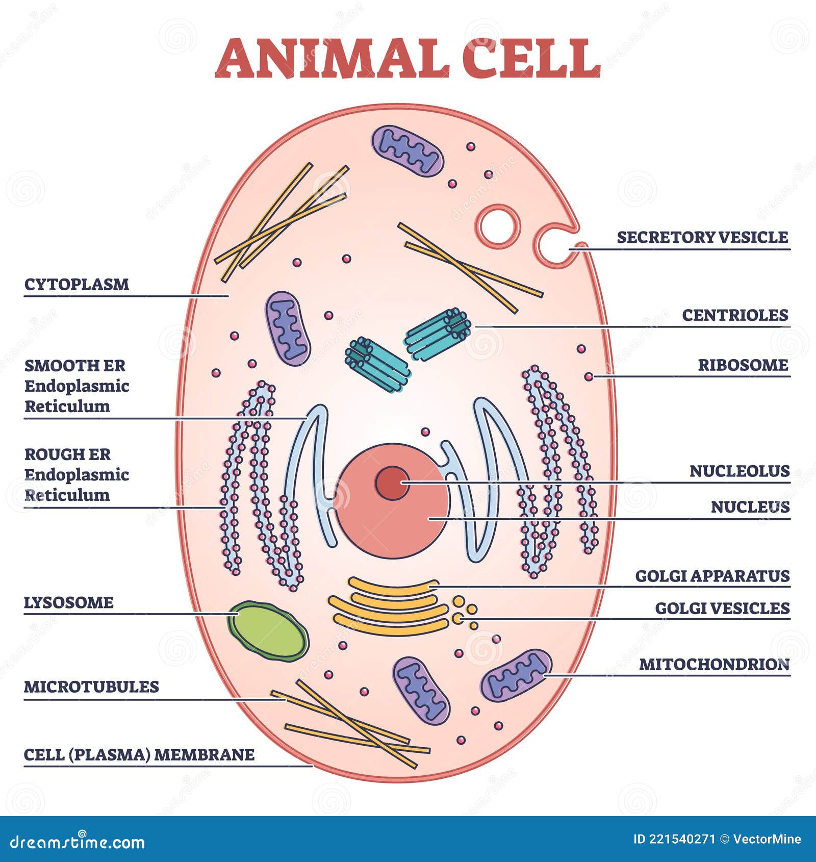
Animal Cell With Labeled Anatomic Structure Parts Diagram Outline Concept Stock Vector Illustration Of Genetic Medical 221540271
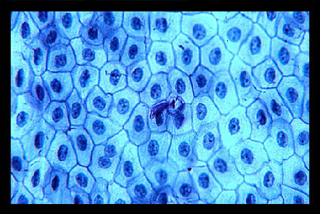
Lab 2 Microscopy And The Study Of Tissues Zoo Lab Uw La Crosse

Draw The Diagram Of An Animal Cell As Seen Through An Electron Microscope And Label The Parts That Brainly In


